Sunday, November 6, 2011

Berlin Medical Historical Museum


In September, I visited Berlin to attend the Council meeting of the European Association of Museums of the History of Medical Sciences at the Berlin Medical Historical Museum. It is a great institution, flourishing under the direction of Thomas Schnalke. We were there to join in a discussion of next year's EAMHMS Congress in Berlin with Thomas. I encourage one and all to attend the 2012 Congress, and in the meantime offer images taken during my visit to the BMHM. Go to Flickr and select my Berlin Medical Historical Museum set.

More pics soon of my visit to Copenhagen and the Medical Museion, after the Berlin trip.

Jim Edmonson

Thursday, September 15, 2011

Sensuous object workshop at the Medical Museion

Later this month I'll be heading to the Medical Museion in Copenhagen for the workshop, the Sensuous Object, in which I'll be examining early stethoscopes. As part of this, I am trying to figure out who first taught and established and codified the protocols of the physical exam. The irony is this: before 1800 doctors hardly touched their patients, instead relying upon the patient's "history" or story of their illness.

This portrait by Winthrop Chandler, of Dr. William Gleason of Connecticut, c.1780, captures the doctor's reticence of physically examining patients. (Image courtesy of the Ohio Historical Society)

Touching was left to surgeons, a more rough and ready lot.


By the end of the century this had all changed and the physical exam was a cornerstone of medical practice, as seen here in images of Sir William Osler examining, palpating, and auscultating a patient, and then contemplating what he had learned. (Images from the Osler Library at McGill University)


A great presentation on how to conduct the exam is given by John Hughes Bennett in Clinical Lecture on the Principles and Practice of Medicine (1844) and an abbreviated version of the same called An Introduction to Clinical Medicine. Bennett credits Leon Rostan, from whom he learned physical examination as a student in Paris, c.1837.

A little digging reveals that Rostan developed his course around 1818 at La Salpetriere and that it was later codified as Traite elementaire de diagnostique, de pronostic, d'indications therapeutiqes ou cours de medecine clinique (1826) and Cours de medecine clinique (1830). Rostan's description of how to conduct a physical exam makes for exceedingly interesting reading.

I'll report back on both what more I uncover, and what I will have learned from the workshop in Copenhagen.


Jim Edmonson

Saturday, July 30, 2011

Hiding in plain view...

While working on the revamp of our gallery of diagnostic instruments, prompted by the generous gift of Don Blaufox's incomparable collection, I went in search of early images of the stethoscope in use. I knew that one of the earliest images appeared in 1819 in the Dictionnaire des sciences medicales. I recalled that Jackie Duffin reproduced this image in her masterful biography of Laennec, To See With a Better Eye (p.212, fig. 9.1). She cited its source as an article on "pectoriloque" by F. V. Merat in the Dictionnaire des sciences medicales vol. 40 (1819).


I checked our online catalog and found we didn't have that dictionary. Struck out, or so I thought. That is, until I found myself in the office of Dzwinka Holian, our library associate director this week. There, on shelving in her office, sat the full 60 volume run of the Dictionnaire des sciences medicales. For some reason it has been uncatalogued, but we're remedying that.

In the meantime, I now have my hands on the original and it's a pretty funny little print. You don't get a sense of that from the image found in Duffin's book. She focused narrowly (understandably) on the scene of the stethoscope, and not the plate in its entirety. The whole plate is full page while the sketch is a very small afterthought, added to what looks like the plate of the stethoscope that appeared in Laennec's treatise on mediate auscultation of 1819. The thing I like best about this whole scenario is that we have a piece of the true cross, as it were. Something from the very dawn of the modern physical examination. And it's not an exact mechanical rendering, but kind of a whimsical take on the physician - patient encounter, with period charm and flavor.


We've got other later period images, lithographs, of percussion and auscultation that appeared in the Western Lancet (Cincinnati, 1850) and have been seldom seen, and we'll be using them in our exhibition, too.


As my friend and colleague Thomas Soderqvist says, exhibits should come from research on collections. When it does, we often more about the wonderful things in our collections than ever suspected...

Jim Edmonson

Friday, May 13, 2011

Juno comes to the Dittrick


Just this past week the Dittrick became the new home to Juno, a transparent woman figure and exhibit mainstay of health museums worldwide. Our Juno lay entombed in her original packing crate, having been shipped to Cleveland from the German Hygiene Museum in Dresden in the early 1950s. Its destination was the Cleveland Health Education Museum (CHEM), which opened in 1940 under the direction of Bruno Gebhardt (formerly of the German Hygiene Museum, 1927-35) and featured state-of-the-art health exhibitry, including Juno.


Jim Edmonson and Linda Spurlock
take stock of the situation.


Franz Tschaikart of Cologne, Germany, crafted Juno upon commission by the German Hygiene Museum. In 1950, a friend of the Health Museum paid $15,000 to bring Juno to Cleveland. "She" first appeared in public on November 13, 1950.


In all, the CHEM housed three Juno-like figures. Over the years they became a Cleveland icon, greeting generations of school kids on field trips to the museum. Sadly, CHEM (later known as HealthSpace Cleveland) closed in 2006 and a vestige of its exhibits and staff came over to the Cleveland Museum of Natural History in 2007. There, as the Department of Human Health headed by Linda Spurlock, they now now offer programming and exhibits to help understand the physiology and frailty of the human body, the sources of disease and the grounding of wellness. Another Juno welcomes museum visitors there.


Our Juno was a backup, but now will greet visitors to the Dittrick as they exit the elevator on the third floor of our home, the Allen Memorial Medical Library. We'll use our Juno to discuss Cleveland's museological heritage, and announce our continuing commitment to showcase issues surrounding women's health.


Jim Edmonson


p.s. - I refer those intrigued by Juno and her male counterparts to Klaus Vogel’s article, “The Transparent Man – Some comments on the history of a symbol,” in Robert Bud, et al, Manifesting Medicine: Bodies and Machines [Artefacts, Studies in the History of Science and Technology , Vol 1], Amsterdam, the Netherlands : Harwood Academic, 1999.

Friday, May 6, 2011

Le Livre Sans Titre, 1830


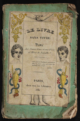
At the recent 2011 meeting of the American Association for the History of Medicine in Philadelphia I picked up a curious tract on the perils of self-abuse, or onanism. Merely speaking the word in polite company invited rebuke in the nineteenth century, so the author simply entitled his work "The Book without a Title," or Le Livre Sans Titre. It's dated 1830 and is distinguished by the charming (may I use that word in this context?) hand-colored depictions of the progressive physical (and moral) decline of the young man in question. A cautionary morality tale indeed! I've translated the captions from French to English, but words are hardly necessary to capture the flavor and tenor of this work...


He was young, handsome; his mother's fond hope


He corrupted himself!... soon he bore the grief of
his error, old before his time... his back hunches
...


A devouring fire sears his gut;
he suffers horrible stomach pains...


See his eyes once so pure, so brilliant;
they are extinguished! a fiery band envelops them.


He can't walk any more... his legs give way


Hideous dreams disturb his slumber...
he cannot sleep
...


His teeth rot and fall out...


His chest burns... he spits up blood...


His hair, once so lovely, falls as if from old age;
his scalp grows bald before his age
....


He hungers; he wants to satiate his appetite;
food won't stay down in his stomach...


His chest collapses... he vomits blood...


Pustules cover his entire body... He is terrible to behold!


A slow fever consumes him, he declines;
all of his body burns up...


His entire body stiffens!... his limbs stop moving...


He is delirious; he stiffens against death;
death gains strength...


At the age of 17, he expires, and in horrible torment


The only library listing for this book in WorldCat is the British Museum. But then again, if you can't give a book a title, it might prove pretty hard to find! We're happy to have it as part of the library for the Percy Skuy Collection at the Dittrick.

Jim Edmonson

Monday, April 11, 2011

Obscura Day at the Dittrick, 2011


Images from the dermatology clinic of William Thomas Corlett (1854-1948), Professor of Dermatology and Syphilology at Western Reserve University, presented by Dittrick's Assistant Curator, Laura Travis, in the Stecher Rare Book Room


We had great fun last Saturday hosting Obscura Day 2011. For a couple of hours we shared some seldom-seen treasures of the Dittrick, ranging from the earliest medical book with woodcut images (The Fasciculus Medicinae of Johannes de Ketham, 1495), to instruments that invoke a grimace and cringe (lithotrites, tonsillotomes, &c), to clinical photos from the 1890s depicting dread diseases like smallpox and syphilis. We're already thinking about Obscura Day 2012 -- how about a magic lantern show, including some medical slides? Sound like fun, eh? For now, here are some more pics from Obscura Day at the Dittrick. It is my understanding that there will be a Flickr page for Obscura Day.

Jim Edmonson


Photos from Dissection: Photographs of a Rite of Passage in American Medicine, 1880-1930 by Warner and Edmonson.


Chief Curator Jim Edmonson presenting a sampler of gems from the rare book collection, on display in the Zverina Room, the Dittrick's seminar room and museum library of trade catalogues.


Jennifer Nieves, Dittrick archivist and museum registrar, showing Jim a selection of instruments from the Dittrick's extensive artifact collection.


Drinks among diaphragms: wine and cheese reception in the history of contraception gallery

Tuesday, April 5, 2011

Dirt: The filthy reality of everyday life (at the Wellcome Collection)

The ravages of cholera, a disease of filth,
as seen in Dirt: the filthy reality of everyday life.


The folks at the Wellcome Collection have done it again. Take a totally banal, prosaic subject -- in this case, dirt -- and re-contextualize it by means of drawings, engravings, film, and artifacts. Though all around us in varying ways, dirt is also taboo, in that talking about it and acknowledging its presence is unsettling. Better off ignoring it, according to common wisdom. But Dirt: The filthy reality of everyday life takes all this head-on. The result is an evocative cultural and social history, driven by visually arresting images. Health and medicine are of course at the heart of this story, but often in surprising and intriguing ways. Can’t wait to see it in person this summer. For the meantime, one can get a better idea of the range and character of Dirt from a great sampler of images and objects on the Wellcome Collection website.


Curatorial credit for the exhibition goes to Kate Forde, James Peto, and Lucy Shanahan.


See mention and reviews of Dirt in the Washington Times, the Guardian, and the Montreal Gazette


Jim Edmonson

Friday, March 18, 2011

CIA students draw surgical artifacts for class


Today we hosted another great group of students from the Biomedical Art program of the Cleveland Institute of Art. Prof. Tom Nowacki brought his medical illustration class to the Dittrick once again to draw surgical instruments from our collections. The sampling varied from O'Dwyer intubation sets to Tarnier's cephalotribe to Bigelow's lithotrite. The assignment revolved around the challenge of showing how these things worked, mechanically, and how they related to the body parts and tissue.


See some of the projects from last year. I'll post those from the 2011 class when they're done in a week or so.


Jim Edmonson